
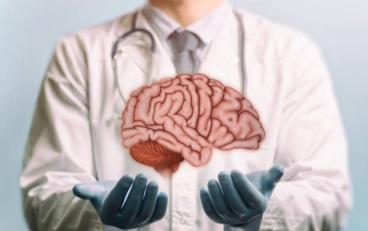
A brain scan alone cannot determine whether you need surgery, a biopsy, monitoring, or another treatment. By the end, you will know what a thorough consultation and brain tumor surgery evaluation should cover, how surgeons define a safe benefit, and which questions help you compare specialists and hospitals.
Key takeaways
- Expect a clinical review, neurological examination, and MRI assessment at the first consultation.
- A complete evaluation covers tumour type, location, symptoms, risks, and your overall health.
- Monitoring, biopsy, or removal depends on tumour behaviour, accessibility, and expected treatment benefit.
- Ask who performs each part of surgery, how risks are managed, and when follow-up occurs.
What happens during the first brain tumor consultation?
The first brain tumor consultation builds a clinical picture before any treatment recommendation. A well experienced neurosurgeon for brain tumor treatment does not decide on surgery from an MRI alone.
1. The surgeon takes a detailed history. Explain headaches, vomiting, seizures, weakness, numbness, speech or vision changes, balance problems, memory difficulties, personality changes, and exactly when each symptom began or worsened.
2. The neurological examination checks strength, sensation, coordination, reflexes, walking, eye movements, language, cognition, and other functions that may be affected by the tumour’s location.
3. Bring every scan and report, including contrast-enhanced MRI images, CT scans, prior treatment records, and pathology. The surgeon also reviews your medicines, seizure history, anticoagulants, antiplatelet drugs, steroid use, and relevant medical conditions.
4. The surgeon then identifies the next clinical question. The same MRI finding can lead to observation, biopsy, surgery, or another treatment in different patients because age, general health, symptoms, previous treatment, tumour size, oedema, growth pattern, blood-vessel relationships, and suspected tumour type change the risk-benefit calculation.
This consultation is not a sales pitch for an operation. Its purpose is to determine what must be clarified next: whether the tumour is causing symptoms, growing, threatening critical brain function, or safe to monitor with repeat imaging.
What does a complete brain tumor surgery evaluation include?
A complete brain tumor surgery evaluation combines symptoms, neurological examination, prior scans, medical history, and specialist review. The surgeon uses this evidence to decide whether treatment is urgent, whether tissue is needed, and whether observation is safer.
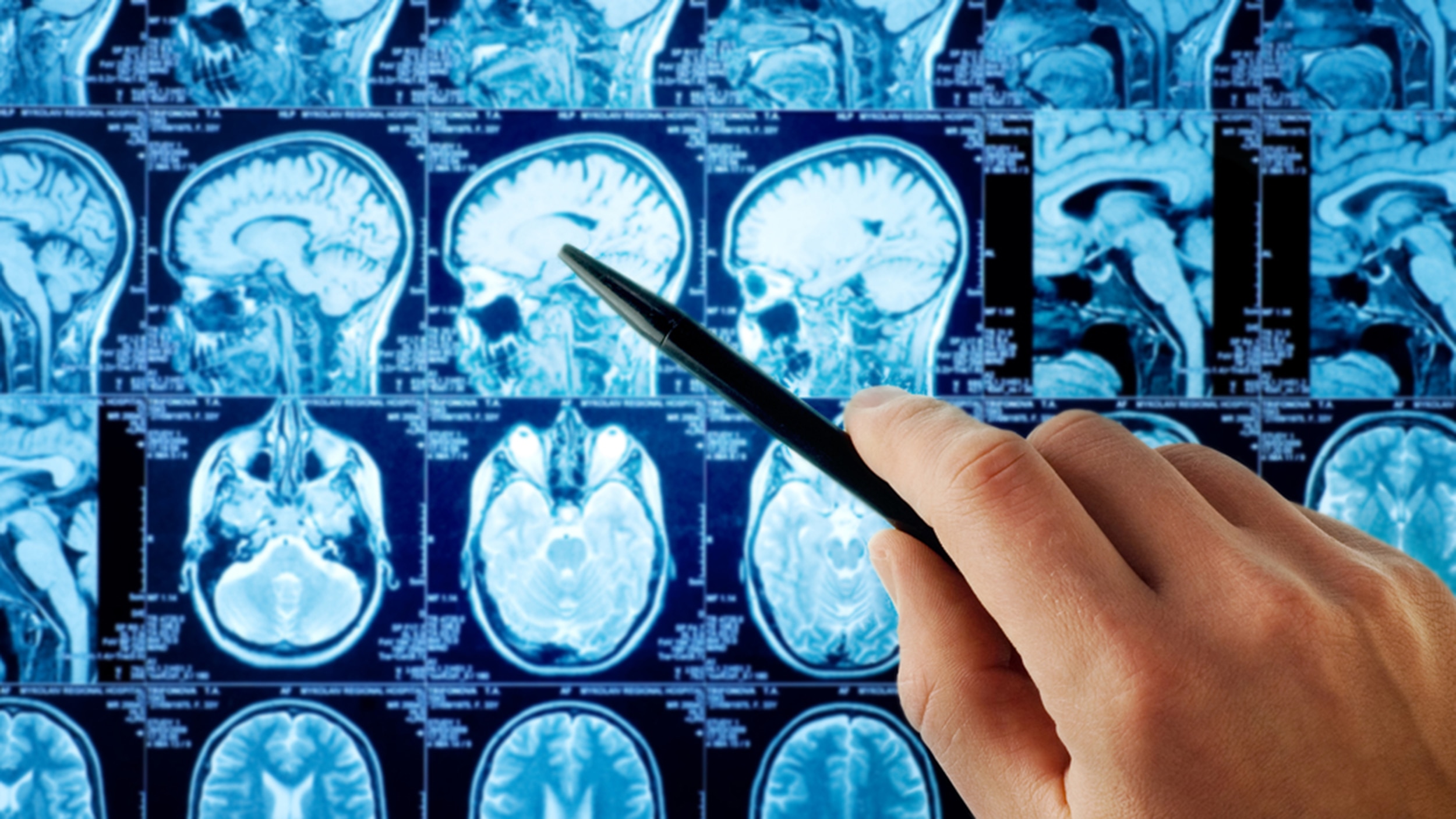
1. Review the contrast-enhanced MRI across all relevant sequences, not one selected image. Assess enhancement, diffusion, haemorrhage, oedema, tumour boundaries, deep structures, and nearby arteries and veins.
2. Add CT when bone involvement, calcification, hydrocephalus, or acute bleeding needs clearer assessment. Consider functional MRI, diffusion-tract imaging, and other mapping tools when the lesion approaches language, movement, vision, memory, or swallowing networks.
3. Complete medical fitness testing with a blood count, clotting tests, kidney function, glucose, ECG, and anaesthesia review. Sellar or skull-base lesions may require endocrine testing; seizures, raised intracranial pressure, hydrocephalus, anticoagulants, steroids, and antiplatelet drugs also affect planning.
4. Identify urgency. Worsening weakness, speech or vision changes, repeated seizures, substantial mass effect, hydrocephalus, haemorrhage, documented growth, or a threatening location increase the case for prompt treatment. A small, asymptomatic, stable lesion may be followed with repeat MRI.
5. Request input from neuroradiology, neuropathology, radiation oncology, or medical oncology when imaging is uncertain, the lesion is unusual, or treatment choices differ. A specialist neuropathology review is valuable when MRI and pathology do not fit.
The final plan also accounts for age, general health, tumour type, and whether you could complete rehabilitation if a neurological deficit occurred.
How does the surgeon choose between monitoring, biopsy, and tumour removal?
A well experienced neurosurgeon for brain tumor treatment does not treat every scan with an operation. The choice depends on symptoms, growth, size, location, suspected tumour type, overall health, and whether treatment risk exceeds the expected benefit.
| Option | Purpose | When it applies |
|---|---|---|
| Observation with serial MRI | Watches for growth or new effects | A small, incidentally found, asymptomatic lesion, such as some meningiomas |
| Stereotactic biopsy | Obtains tissue without attempting full removal | A deep, diffuse, inaccessible, or high-risk lesion |
| Maximal safe resection | Removes as much tumour as possible while protecting function | A suspected glioma or other tumour where resection offers meaningful benefit |
| Debulking or diversion | Reduces pressure or treats a complication | A large mass causing pressure, hydrocephalus, or another urgent problem |
A biopsy is diagnostic surgery, not an attempt to remove the tumour. Tissue supports an integrated WHO CNS diagnosis and molecular testing, including IDH mutation, 1p/19q codeletion, ATRX status, and relevant histone alterations; the report can remain incomplete until these results return.
For a suspected glioma, maximal safe resection means stopping before removal creates an unreasonable risk to speech, movement, vision, memory, swallowing, or another critical function. A visible remnant can be safer than aggressive removal.
Treatment may instead use radiotherapy, chemotherapy, targeted treatment, or a combination, selected according to pathology, molecular findings, growth, symptoms, location, and health.
How does an experienced brain surgeon make tumour removal safer?
Safer tumour removal starts with choosing a corridor around functional and vascular anatomy, not simply the shortest route to the MRI target. An experienced brain surgeon weighs the expected benefit against possible harm to speech, movement, vision, memory, swallowing, or other critical functions.
Planning may include:
- Neuronavigation to track the tumour and planned corridor
- Functional MRI and diffusion-tract imaging to locate important networks and fibre pathways
- Intraoperative ultrasound or MRI to update the tumour’s position during surgery
- Cortical or subcortical stimulation mapping and motor or sensory pathway monitoring
| Surgical plan | Main purpose | Why it may be chosen |
|---|---|---|
| Maximal safe resection | Remove as much tumour as safely possible | The tumour is accessible without an unreasonable functional risk |
| Limited resection or biopsy | Obtain tissue or reduce pressure while preserving function | The lesion lies near the motor cortex, language network, optic pathways, brainstem, or major vessels |
| Staged or nonsurgical treatment | Control disease without immediate extensive removal | Surgery carries greater risk than observation, radiation, or systemic treatment |
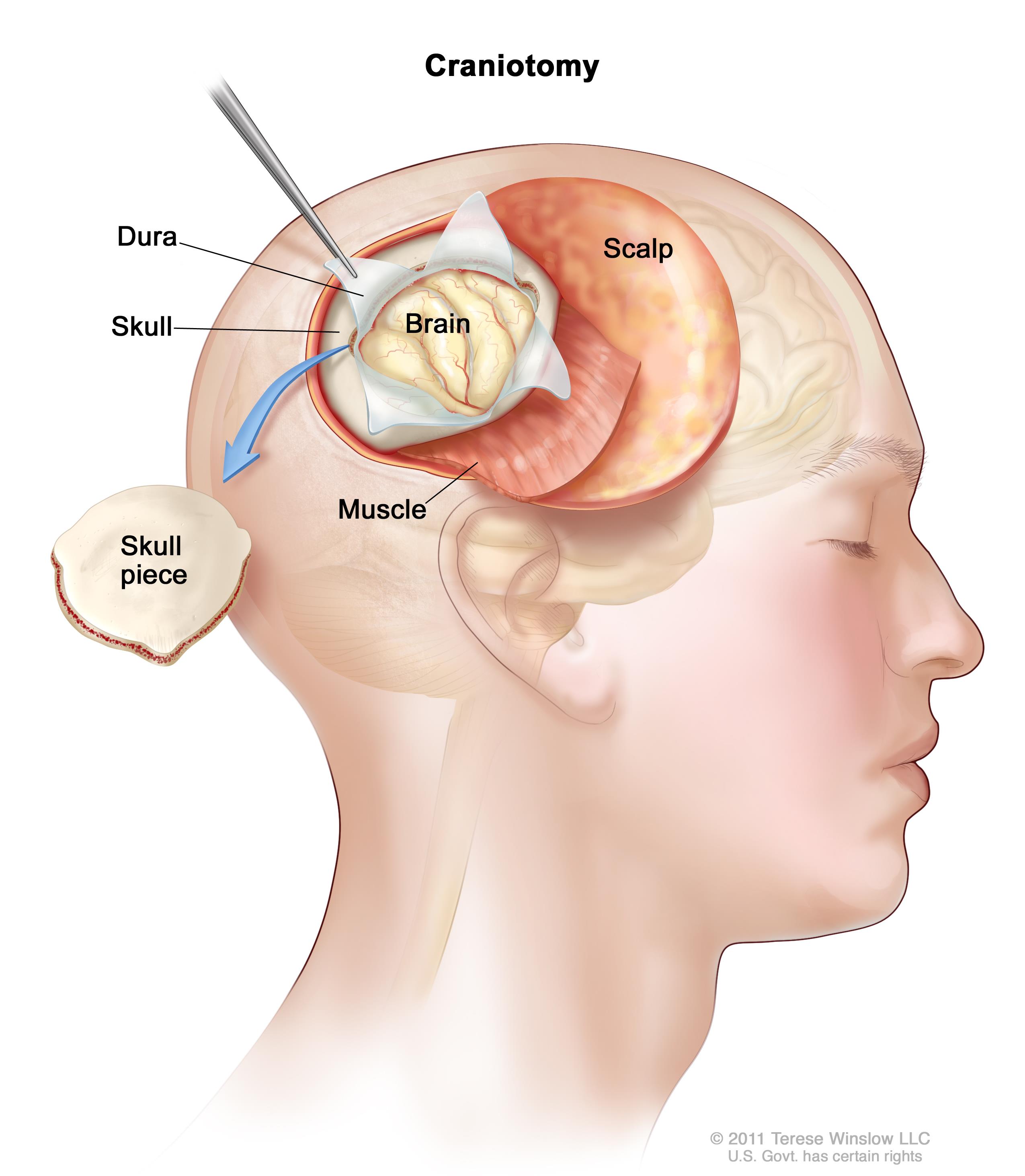
Awake craniotomy is a selective option for tumours near language or other functions that can be tested during surgery. It requires patient cooperation, suitable anaesthesia planning, and an assessment that the patient can participate; it is not automatically safer or more advanced.
Before consent, ask which benefit is expected, whether incomplete removal is safer, and which deficits could be temporary or permanent. Discuss bleeding, infection, seizures, swelling, stroke, cerebrospinal-fluid leakage, venous thromboembolism, and recurrence. A single complication percentage cannot represent every tumour location or operation.
Compare experience with your tumour and planned procedure, not labels such as “best” or “leading” alone.
What should you ask about the team, recovery, and follow-up?
Before choosing a brain tumor neurosurgeon in Pune, ask who will review your MRI, pathology, and treatment plan. Find out whether a specialist neuropathologist can provide a second opinion, which mapping and monitoring tools are available, and how the proposed benefit will be measured.
Ask these questions:
- Will neuroradiology review the scans, and will functional MRI, diffusion-tract imaging, neuronavigation, or stimulation mapping be used?
- What function could the operation affect, and what alternatives exist?
- Are neuroanaesthesia, neurocritical care, oncology, and rehabilitation available through one care pathway?
- Who will manage seizures, steroids, neurological changes, and rehabilitation after discharge?
A second opinion is particularly useful when the diagnosis is unusual, MRI and pathology conflict, the lesion lies in an eloquent region, or the operation carries high risk. It can clarify the diagnosis without implying that the first surgeon made an error.
| Question | What to confirm | Why it matters |
|---|---|---|
| Early MRI | When the postoperative scan will occur | Shows the initial extent of removal |
| Integrated pathology | When molecular results will be complete | Guides further treatment |
| Recovery plan | Seizure, steroid, and rehabilitation management | Detects problems early |
| Follow-up MRI | Timing based on tumour biology and treatment | Avoids one fixed schedule for every tumour |
Postoperative care is part of treatment. The team reviews the early MRI, confirms integrated pathology, monitors neurological recovery, and coordinates radiotherapy or systemic treatment when indicated. For someone assessing Dr Dilip Kiyawat in Pune and Pimpri-Chinchwad, this checklist tests team coordination rather than reputation alone.
Related items
 | Brain Surgery ₹1200 Brain tumours are abnormal growths of cells within the brain or the surrounding structures. View service → |
Brain Surgeon Here are numerous types of brain surgery. The type used is based on the area of the brain and the condition being treated. View product → |
Frequently asked questions
What happens during the first brain tumor consultation?
The neurosurgeon reviews your symptoms, medical history, scans, previous reports, and neurological examination before discussing treatment options.
What does a complete brain tumor surgery evaluation include?
It assesses the tumour’s type, size, location, growth pattern, relationship to critical brain structures, surgical risks, and your general health.
How does the surgeon choose between monitoring, biopsy, and tumour removal?
The choice depends on the scan findings, suspected tumour type, growth or symptoms, accessibility, and whether tissue diagnosis or removal offers greater benefit.
How does an experienced brain surgeon make tumour removal safer?
The team maps critical brain functions, plans the surgical route, reviews anaesthesia and monitoring needs, and prepares for risks specific to the tumour’s location.
What should you ask about the team, recovery, and follow-up?
Ask who will perform the surgery, expected hospital stay, possible complications, rehabilitation needs, pathology reporting, follow-up scans, and warning signs after discharge.
Related items
| TUMOR & CANCERS Brain Tumor & Cancer An Cerebral aneurysm or intracranial aneurysm is when the internal walls of the artery through which the blood flows in the brain becomes thin and... |
 | Types of brain tumours- Primary brain tumors are tumors that originate in the brain itself, as opposed to secondary tumors that spread to the brain... View service → |