
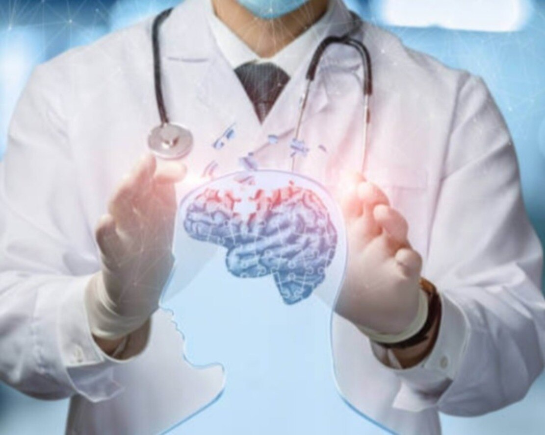
A strong surgical consultation explains why an operation is needed, what it is intended to achieve, and how the team will protect speech, movement, vision, memory and other critical functions. By the end, you will know which questions to ask, how to compare surgeons and hospitals, what happens in the operating room, and what recovery involves.
Key takeaways
- Ask the surgeon to review your MRI or CT images with you.
- Compare surgical experience, hospital facilities, intensive care, and rehabilitation support.
- Confirm the surgery’s goal, tumour location, functional risks, and backup treatment plan.
- Before discharge, get medication, wound-care, warning signs, and follow-up instructions in writing.
What should a brain tumor surgeon consultation establish?
A brain tumor surgeon consultation should establish the treatment goal and whether its expected benefit outweighs the risks. The surgeon should review the actual MRI or CT images, not only the radiology report, then relate the tumour’s size, depth and location to your symptoms, neurological examination and functional priorities.
Bring details of previous cancer, seizures, medicines, blood thinners and earlier scans.
Ask the surgeon to explain these points:
- Which symptoms result from the tumour, swelling or hydrocephalus, and which brain functions lie near it?
- What could happen without treatment, and how would observation be monitored?
- What are the expected benefits and specific risks of surgery, including weakness, speech or vision changes, seizures, bleeding, infection, swelling and cerebrospinal-fluid leak?
- Is the safest procedure a stereotactic biopsy, resection, pressure-relieving operation or treatment for hydrocephalus?
- How will tissue be tested? A visual impression or frozen section does not settle every diagnosis; final pathology can combine histology with IDH, 1p/19q, ATRX, TP53 and MGMT findings.
- What further treatment could follow, including radiation or chemotherapy?
| Option | Main purpose | Important planning point |
|---|---|---|
| Observation | Monitor a stable or lower-risk finding | Requires scheduled scans and symptom review |
| Biopsy | Obtain tissue diagnosis | Useful when the tumour is deep, diffuse or near critical pathways |
| Resection | Remove as much as safely possible | The goal is maximal safe resection, not removal at any cost |
| Radiation or chemotherapy | Treat remaining or unsuitable tumour | Selection depends on pathology, molecular results and overall health |
A clear consultation also explains whether surgery aims at tissue diagnosis, maximal safe resection, pressure relief or hydrocephalus treatment.alc 金尊
How can you judge the best neurosurgeon and hospital?
The best neurosurgeon during brain tumor surgery is not identified by a city label or a “best” claim. Look for experience with your tumour type and its location, a meaningful case volume, and results reported as complications, extent of removal, neurological outcomes and readmissions.
Ask these questions before choosing:
- Who will perform the operation, and how many similar tumours in this location have they treated?
- Can the team offer stereotactic biopsy, resection or pressure-relieving surgery when each is appropriate?
- How will the sample be handled for histology and molecular tissue testing, including IDH, 1p/19q, ATRX, TP53 and MGMT when relevant?
- Is awake mapping available for a tumour near language or movement networks, and is intraoperative ultrasound, MRI or updated neuronavigation available when brain shift reduces navigation accuracy?
- Does the hospital provide 24-hour neuro-anaesthesia, neuro-ICU care and emergency reoperation?
- Are neuroradiology, neuropathology, radiation oncology, medical oncology and rehabilitation coordinated under one treatment plan?
For brain surgery in Pune, these answers matter more than the location on the hospital sign. A surgeon such as Dr Dilip Kiyawat should be assessed through this same evidence-based checklist, not reputation alone. Ask who will manage a sudden neurological change overnight and who will coordinate radiotherapy, oncology and rehabilitation after discharge.
A capable surgeon without round-the-clock hospital support leaves gaps precisely when complications demand rapid action.
Related items
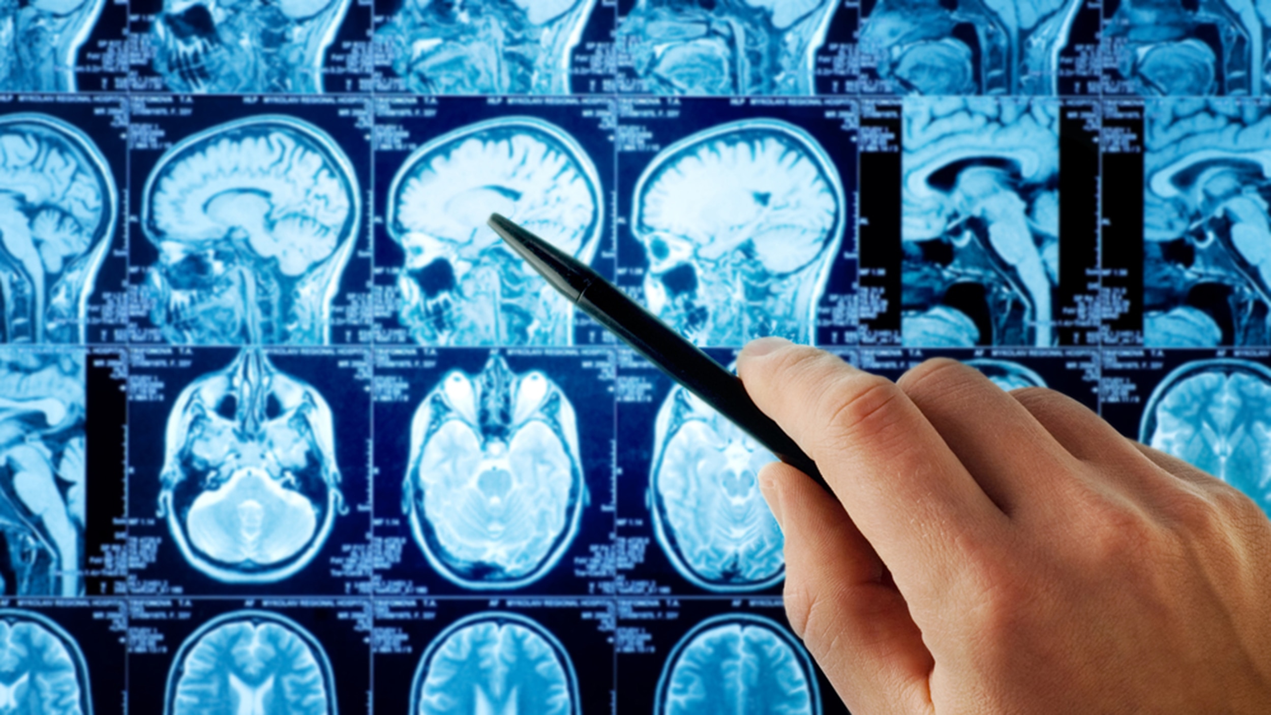 | Brain Surgery ₹1200 Brain tumours are abnormal growths of cells within the brain or the surrounding structures. View service → |
Brain Surgeon Here are numerous types of brain surgery. The type used is based on the area of the brain and the condition being treated. View product → |
What happens during the brain tumor surgery process?
The brain tumor surgery process starts with preoperative clearance, updated MRI or CT review, blood tests, medicine and blood-thinner instructions, and hospital admission. The team confirms whether the goal is tissue diagnosis, maximal safe removal, pressure relief, or treatment of hydrocephalus.
1. Anaesthesia is given before you are positioned, usually with your head secured in a frame. The team clips or shaves only the hair needed for the incision, cleans the skin, and plans either a craniotomy or a minimally invasive route.
Endoscopic surgery may reach selected ventricular or skull-base tumours through a small opening; a pressure-relieving drain or shunt may be used instead of tumour removal.
2. The surgeon may perform a gross total resection, removing all visible tumour; a subtotal resection, leaving visible tumour to protect critical tissue; or debulking, reducing tumour volume and pressure without attempting complete removal. A stereotactic biopsy takes small samples through a precisely planned needle, which suits some deep, diffuse, or functionally dangerous tumours.
3. Neuronavigation guides the route, while updated MRI or intraoperative ultrasound checks the changing anatomy. Brain shift after skull opening, cerebrospinal-fluid release, or tumour removal makes the original map imperfect. Electrophysiological monitoring, cortical and subcortical mapping, diffusion-tract planning, functional MRI, and suitable 5-ALA fluorescence add information, but none guarantees preservation.
Awake craniotomy is reserved for selected tumours near language or movement networks. During mapping, you perform naming, counting, or movement tasks while stimulation identifies tissue to preserve. After removal or biopsy, the surgeon closes the wound and transfers you to recovery or neuro-ICU for neurological observation.
How do tumour location, surgical goals and risks shape the result?
The result is shaped by tumour location, the purpose of surgery and the disability risk of reaching it. The usual goal is maximal safe resection, not automatic removal of every visible abnormality.
A tumour beside language, movement, vision, memory, breathing or swallowing pathways, or major white-matter tracts, may be deliberately left partly behind if further removal could cause permanent loss of function.
Ask the surgeon to connect each risk to your tumour, health and planned procedure:
- Bleeding, infection or seizures after surgery.
- Swelling, stroke or cerebrospinal-fluid leak.
- Blood clots, anaesthesia complications or problems caused by other medical conditions.
- New weakness, speech or vision changes, or cognitive effects.
- Incomplete removal, the need for additional treatment and tumour recurrence.
The surgeon’s visual impression does not establish the final diagnosis. Pathology combines the surgical sample, histology and molecular findings, which can include IDH mutation, 1p/19q codeletion, ATRX, TP53 and MGMT promoter methylation. The report may take longer than an initial frozen section and can change decisions about radiation, chemotherapy or surveillance.
Ask which functions are at risk, what benefit further removal offers, and how the team will decide when stopping is safer than taking more tissue.
What happens immediately after surgery and before discharge?
After surgery, you may spend the first night in a neuro-ICU or ward, depending on the operation and your condition. Staff repeatedly check alertness, pupils, speech, strength, sensation and coordination, alongside blood pressure, breathing and temperature.
They treat pain and nausea, monitor swelling and sodium changes, and watch for seizures, infection, bleeding, venous thrombosis and cerebrospinal-fluid leakage.
1. Mobilisation starts when safe: sitting, standing and walking with nursing or physiotherapy support. Physiotherapy addresses weakness and balance; speech therapy assesses communication or swallowing, while occupational therapy helps with daily tasks when needed.
2. An MRI or CT is usually performed within 24–48 hours after resection. This early image records postoperative anatomy and any residual enhancing tumour as a baseline; later reactive enhancement can obscure that distinction. It does not alone decide whether you need radiotherapy or chemotherapy.
3. The pathology report combines histology with molecular results to identify the tumour and guide further treatment. Ask when the report is expected and how it will change follow-up.
Steroids reduce tumour-related swelling but can cause high blood sugar, insomnia, mood changes, infection risk and muscle weakness, so clinicians use the lowest effective dose and taper when safe. Anti-seizure medicine is not automatic for a seizure-free patient; a prior seizure or another indication changes that decision.
Discharge depends on stable neurological findings, controlled symptoms, safe walking or a rehabilitation plan, adequate eating and drinking, and a wound without concerning leakage or infection. Follow-up covers wound review, pathology, imaging, rehabilitation and warning signs needing urgent assessment.
Frequently asked questions
What should a brain tumor surgeon consultation establish?
It should establish the treatment goal, expected benefit, major risks, and whether surgery is appropriate after reviewing your MRI or CT images, symptoms, examination, and priorities.
How can you judge the best neurosurgeon and hospital?
Compare the surgeon’s experience with your tumour type and location, then assess the hospital’s imaging, operating-room, intensive-care, pathology, rehabilitation, and emergency support.
What happens during the brain tumor surgery process?
The process includes preoperative assessment, anaesthesia, positioning, tumour access and removal or biopsy, tissue analysis, recovery monitoring, and planning for further treatment.
How do tumour location, surgical goals and risks shape the result?
Tumour depth and proximity to speech, movement, vision, memory, or other critical areas influence whether the goal is complete removal, partial removal, biopsy, or symptom relief.
What happens immediately after surgery and before discharge?
The team monitors consciousness, movement, speech, pain, seizures, wound healing, and scans, then provides medication, activity, warning-sign, and follow-up instructions before discharge.
Related items
| TUMOR & CANCERS Brain Tumor & Cancer An Cerebral aneurysm or intracranial aneurysm is when the internal walls of the artery through which the blood flows in the brain becomes thin and... |
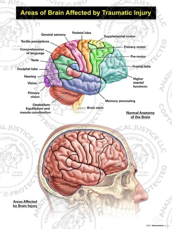 | Treatment of Head injury ₹1200 Management of head injuries 1. Extensive scalp laceration- . The bleeding is controlled by suturing the scalp. . View service → |
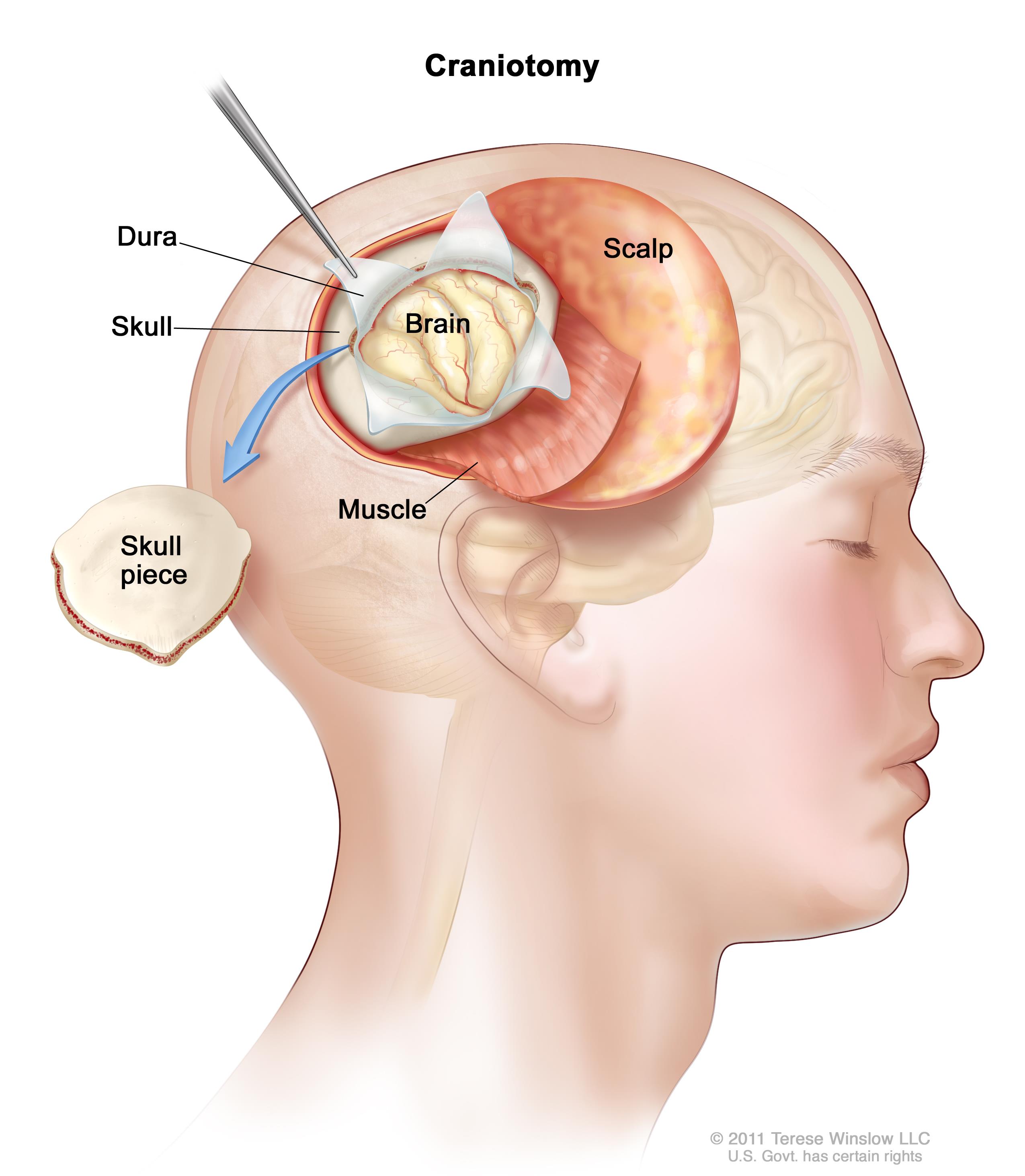 | Types of brain tumours- Primary brain tumors are tumors that originate in the brain itself, as opposed to secondary tumors that spread to the brain... View service → |
 | Spine Surgery Spine Surgeon in Pune Dr. Dilip S. Kiyawat works as Neurosurgeon at Jehangir Hospital, Pune, Maharashtra, India. He completed his M.Ch.(Neuro). View product → |