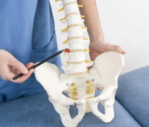


A vertebral fracture caused by osteoporosis is not automatically a reason for surgery. You need to know whether the fracture is new, whether it is stable, whether nerves are affected, and how poor bone quality changes both the operation and recovery plan.
Key takeaways
- Sudden back pain after minor strain can indicate a vertebral compression fracture.
- MRI helps identify a recent fracture and rule out nerve or spinal cord pressure.
- Stable fractures often need pain control, osteoporosis treatment, and a fitted brace.
- Ask about bone-strengthening treatment, fixation options, complications, and follow-up.
What an Osteoporotic Vertebral Fracture Looks and Feels Like
Osteoporosis thins the vertebra’s internal, honeycomb-like bone until it cannot withstand normal loading. A minor fall, lifting movement, sudden twist, or coughing episode can then crack the vertebral body. Pain is usually sudden, deep, and centred in the back; standing, walking, turning, or coughing can intensify it.
Some fractures cause height loss or a gradually rounded posture.
| Pattern | What happens | How it may appear or feel |
|---|---|---|
| Compression | The vertebral body is squeezed down | Localised back pain, often without nerve symptoms |
| Wedge | The front collapses more than the back | Forward stooping and pain when standing upright |
| Burst | The body breaks in multiple directions, sometimes pushing fragments backward | Severe pain with possible spinal-canal or nerve involvement |
| Collapse | A weakened vertebra progressively loses height | Increasing deformity, persistent pain, or repeated collapse |
Seek urgent assessment for new leg weakness, numbness, loss of bladder or bowel control, inability to stand, or severe unremitting pain. Fever after a procedure also needs prompt review. These symptoms can indicate nerve compression, infection, or another serious cause rather than uncomplicated osteoporotic fractures.
Atypical pain, fever, or unexplained collapse requires MRI to distinguish osteoporosis from infection or cancer; an X-ray finding alone does not automatically mean surgery.
How Doctors Confirm a New Fracture and Find Its Cause
Doctors confirm a new fracture by matching your pain, examination, and imaging rather than relying on an X-ray alone. The clinician records the trigger, pain location, duration, fever, cancer history, steroid use, previous fractures, and symptoms in the legs, bladder, or bowel, then checks strength, sensation, reflexes, walking, and spinal tenderness.
The diagnostic pathway usually includes:
- Standing or lying X-rays to show vertebral height loss, wedge shape, alignment, and progressive collapse.
- MRI to identify bone-marrow oedema, which supports a recent fracture, and to detect nerve compression, infection, marrow replacement, or malignant collapse.
- CT to define a burst pattern, posterior-wall break, retropulsion into the spinal canal, or unstable bone fragments.
- DEXA scanning and bone-quality assessment to establish osteoporosis; these tests do not prove that the painful collapse is new.
- Blood tests such as a complete blood count, C-reactive protein, erythrocyte sedimentation rate, calcium, alkaline phosphatase, and kidney-function tests when infection, metabolic disease, or cancer is possible.
An old collapse usually lacks acute marrow oedema and may match earlier images, while a new fracture produces pain that correlates with the imaging finding. A compression fracture alone does not justify surgery. Neurological deterioration, canal compromise with matching symptoms, instability, progressive deformity, or disabling pain despite properly conducted nonoperative care changes the discussion.
A decision about osteoporotic spine fracture surgery in Pune should rest on those findings, not on a label such as “experienced” alone.
When Treatment Is Bracing, Medication, Cement, or Open Surgery
A fracture without neurological deficit, major instability, or worsening deformity is usually treated without surgery first. Analgesia, early mobilisation, a short period of bracing, and osteoporosis treatment often control pain while avoiding the harms of bed rest, including thrombosis, pressure injuries, respiratory problems, and loss of strength.
| Option | What it involves | When it applies |
|---|---|---|
| Analgesia and mobilisation | Pain relief followed by safe walking | Stable fractures without nerve compression |
| Brace | Temporary external support | Painful movement during early healing; prolonged use weakens muscles |
| Osteoporosis treatment | Calcium and vitamin D review, fall prevention, and prescribed bone medicine | Every confirmed fragility fracture, whether or not surgery occurs |
| Vertebroplasty | Cement injection into the collapsed vertebral body | Selected persistent, fracture-related pain after nonoperative care; routine benefit remains uncertain |
| Kyphoplasty | Balloon cavity creation before cement injection | Selected painful collapse when height restoration or controlled cement placement matters |
| Decompression and fixation | Removal of nerve pressure, stabilisation with implants, or both | Progressive neurological deficit, canal compromise, major instability, or deformity |
Cement does not cure osteoporosis, and open surgery carries greater anaesthetic, wound, implant, and neurological risks in fragile bone. Avoiding surgery is safer when pain is improving, alignment is acceptable, and neurological function remains intact.
A sound spine fracture surgery process documents fracture-related pain, failed conservative treatment, bone quality, medical fitness, and the reason each alternative was rejected.
The Spine Fracture Surgery Process From Admission to Discharge
Admission starts with a neurological examination: leg strength, sensation, reflexes, walking ability, and bladder or bowel symptoms are documented. Any deterioration or cord and nerve compression can change the urgency of treatment.
- Review X-rays, CT, and MRI to confirm fracture age, spinal-canal involvement, instability, deformity, and pain correlation. Standing alignment films are obtained when safe.
- Optimise medical risks before surgery. The team reviews anaemia, infection, kidney function, calcium and vitamin D status, diabetes, blood-thinning medicines, bone quality, and heart or lung disease.
- Plan anaesthesia around frailty, breathing risk, medicines, positioning, and the expected duration of surgery. The anaesthetist explains fasting, airway management, and postoperative pain control.
- Select the procedure that matches the fracture and neurological findings. Options include cement augmentation, decompression, fixation, or a combination; an X-ray showing compression alone does not automatically justify surgery.
- Monitor nerve function, blood loss, blood pressure, oxygen levels, and wound condition during and after the operation. New weakness, worsening numbness, fever, or severe pain needs immediate review.
- Begin pain-controlled mobilisation with physiotherapy. The team checks walking safety, brace requirements, clot risk, and the ability to manage basic activities.
- Discharge instructions cover wound care, medicines, lifting limits, warning symptoms, and osteoporosis treatment. Follow-up includes clinical review and scheduled imaging to detect infection, construct failure, recurrent collapse, or another fracture.
This spine fracture surgery process applies whether you are arranging care locally or comparing osteoporotic spine fracture surgery in Pune. Recovery continues after discharge through rehabilitation and secondary-fracture prevention.
How Poor Bone Quality Changes Surgical Risk and Recovery
A screw relies on firm bone to resist pullout. In osteoporotic fractures, reduced bone density gives the threads less purchase, so screws can loosen, migrate, or fail under spinal load. Weak vertebrae also increase the risk of further collapse around a construct.
The fixation plan is tailored to bone quality, fracture stability, alignment, and nerve compression. Options may include:
- Extending fixation across more vertebrae to spread the load
- Using larger screws or different screw trajectories
- Adding cement augmentation to selected screws
- Combining fixation with decompression when bone or fracture fragments compress nerves
Cement improves screw purchase but introduces its own hazard. Leakage into the spinal canal can injure a nerve, while leakage into veins can affect the lungs. Surgeons reduce this risk through careful imaging, controlled cement volume, suitable screw selection, and monitoring during injection.
Recovery continues after discharge. Neurological checks look for new weakness or numbness; physiotherapy rebuilds safe movement and strength; imaging and clinical reviews identify loosening, recurrent collapse, infection, or an adjacent-level fracture.
Another fracture-prevention plan is essential. It may include prescribed osteoporosis medication, adequate calcium and vitamin D, fall-proofing the home, and weight-bearing activity when safe. Without bone treatment, technically successful surgery does not remove the risk of another vertebral or hip fracture.
Questions to Ask When Comparing a Spine Surgeon in Pune
A search for “well experienced neurosurgeon spine surgery for process osteoporotic fractures in pune” is not a clinical assessment. Ask an experienced spine surgeon in Pune to link each recommendation to your examination, MRI or CT findings, and the risks of waiting.
- What neurological deficit, instability, progressive deformity, or fracture finding makes surgery safer than analgesia, mobilisation, bracing, and osteoporosis treatment?
- How will poor bone quality change fixation: longer constructs, cement-augmented screws, or another strategy? How will you limit cement leakage and nerve injury?
- Why is vertebroplasty, kyphoplasty, decompression, fixation, or nonoperative care suitable, and what alternatives were rejected?
- Who will coordinate calcium and vitamin D review, anti-osteoporosis medication, renal or metabolic checks, imaging, physiotherapy, and follow-up?
When consulting Dr Dilip Kiyawat, ask who personally reviews your images and neurological findings, performs the operation, and remains responsible after discharge.
Related services
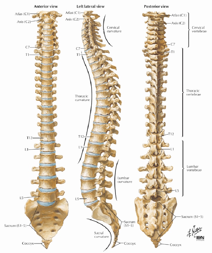 | Spine Surgery Dr. Dilip S. Kiyawat has wide experience in treating spinal disorders. View service → |
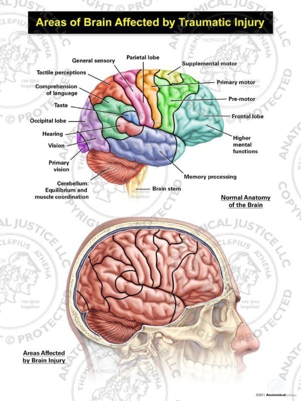 | Treatment of Head injury ₹1200 Management of head injuries 1. Extensive scalp laceration- . The bleeding is controlled by suturing the scalp. . View service → |
Frequently asked questions
What does an osteoporotic vertebral fracture feel like?
It usually causes sudden, deep pain in the middle or lower back after a minor fall, lift, twist, cough, or sometimes no clear injury. Standing, walking, turning, and coughing can worsen the pain.
How do doctors confirm a new osteoporotic spine fracture?
Doctors use your symptoms, examination, and imaging. X-rays can show vertebral collapse, while MRI can identify bone marrow swelling from a recent fracture and reveal nerve or spinal cord compression. CT provides detailed views of the bone and may help plan surgery.
When is surgery needed for an osteoporotic spine fracture?
Treatment depends on pain, spinal stability, deformity, neurological symptoms, and response to non-surgical care. Bracing and medication often suit stable fractures. Cement augmentation or fixation may be considered for persistent pain, instability, deformity, or nerve compression.
What happens during spine fracture surgery?
After admission and preoperative assessment, the team reviews imaging, medicines, anaesthesia risks, and bone quality. Surgery may involve cement injection, screws and rods, decompression, or a combination. After monitoring, pain control, mobilisation, and wound checks, discharge planning begins.
How does osteoporosis affect spine surgery recovery?
Poor bone quality reduces screw holding strength and increases risks such as implant loosening, adjacent fractures, delayed healing, and loss of spinal alignment. Surgeons may alter fixation, use bone-strengthening treatment, and arrange closer imaging and follow-up.