
Brain tumor surgery can mean a needle biopsy, partial or complete removal, pressure relief, or treatment for blocked cerebrospinal fluid—not one standard operation. By the end, you will know when surgery is considered, how doctors choose an approach, what tests and risks matter, and how to prepare for a useful consultation.
Key takeaways
- MRI findings, symptoms, seizures, and neurological signs guide the first surgical discussion.
- Surgery is not always the first choice; observation, biopsy, radiation, or medication may fit the diagnosis.
- Biopsy identifies tumor type when full removal is unsafe or unnecessary.
- Pathology after surgery determines further treatment, follow-up scans, and specialist referrals.
What happens from the first consultation through recovery?
1. The first consultation includes a neurological examination and a review of when symptoms began, how they changed, and whether seizures occurred. The surgeon assesses MRI scans, often with and without contrast, then discusses whether tissue diagnosis is needed and what the operation should achieve.
2. Brain tumor surgery explained in plain language means choosing the safest useful procedure: a stereotactic needle biopsy for tissue, resection to remove accessible tumor, debulking to reduce pressure, decompression, or shunt placement for hydrocephalus. Complete removal is not always possible or curative, especially when tumor cells infiltrate normal-looking brain.
3. If surgery proceeds, anesthesia keeps you unconscious and pain-free while the team monitors vital signs and neurological function in hospital. Pathology identifies the tumor type and grade, with molecular testing when indicated. A postoperative MRI checks residual tumor, swelling, bleeding, or other changes.
4. Recovery depends on the operation and the brain region treated. Your plan covers wound care, seizure treatment, weakness, speech, vision, memory, mobility, and rehabilitation, followed by surveillance MRI and any needed radiotherapy, chemotherapy, targeted treatment, or observation. Ask when to resume medicines and activity, and which symptoms require urgent contact.
When is brain tumor surgery needed, and when is it not the first choice?
When brain tumor surgery is needed depends on symptoms, scan changes, diagnostic uncertainty, and the risk of reaching the lesion. An operation is more likely when the tumor causes a problem that needs relief or tissue diagnosis:
- Progressive weakness, numbness, speech difficulty, vision loss, personality change, or other neurological deficits
- Seizures that require treatment or a tissue diagnosis
- Pressure or mass effect causing headaches, vomiting, drowsiness, or worsening brain function
- Bleeding into or around the lesion
- Hydrocephalus, when blocked cerebrospinal-fluid flow enlarges the brain’s fluid spaces
- Significant growth on serial MRI
- Uncertainty about the diagnosis when tissue will change treatment
- A lesion that can be treated with acceptable risk through biopsy, removal, decompression, or another procedure
Seek emergency assessment, not a routine appointment, for a first seizure, rapidly worsening weakness or speech, reduced consciousness, repeated vomiting with severe headache, or sudden neurological deterioration.
Surgery is not automatically the best first choice. A small, incidentally discovered, slow-growing, or asymptomatic lesion may be monitored with repeat MRI. Radiotherapy, chemotherapy, targeted treatment, or supportive care may fit better when the tumor responds to those treatments, surgery carries substantial neurological risk, or the patient’s overall condition makes an operation unsafe.
How do surgeons choose between biopsy, removal, and specialised approaches?
Biopsy and resection answer different questions. A biopsy obtains tissue for pathology and molecular testing; resection removes as much tumour as safely possible. In plain language, brain tumour surgery explained means matching the objective to the tumour and the brain around it, not chasing complete removal at any cost.
| Option | What it does | When it applies |
|---|---|---|
| Stereotactic biopsy | Uses a needle to obtain tissue through a small opening | When diagnosis is needed but removal carries high risk |
| Craniotomy with resection or debulking | Removes all visible tumour, or reduces its volume | When safe access is possible; subtotal resection is safer near language, movement, vision, memory, or deep structures |
| Endoscopic surgery | Uses a camera and narrow instruments | For selected ventricular, skull-base, or fluid-blocking lesions |
| Decompression | Relieves pressure without necessarily removing the tumour | When swelling or mass effect threatens brain function |
| Shunt placement | Diverts cerebrospinal fluid | When hydrocephalus results from blockage |
| Awake craniotomy | Tests speech or movement during selected operating stages | When mapping critical functional areas could protect them |
Planning combines neurological examination; MRI with and without contrast; CT; functional MRI; diffusion-tract imaging or tractography; PET; and angiography. Neuronavigation, intraoperative ultrasound or MRI, neurophysiological monitoring, and awake mapping guide the operation, but none removes the risk of deficit, bleeding, seizures, or residual tumour.
What does an experienced brain tumor surgeon evaluate beyond years of practice?
An experienced brain tumor surgeon evaluates the whole clinical picture, not just years in practice. A neurosurgeon with above 40 years of experience in brain tumor surgery may bring valuable judgement, but years alone do not prove current expertise in a particular operation.
1. Review location, size, growth pattern, contrast enhancement, edema, bleeding, hydrocephalus, suspected pathology, and molecular markers. Match these findings with symptoms, seizures, age, general health, anticoagulants and other medicines, functional priorities, and the risk of neurological deficit.
2. Set the safest objective. Safe resection is preferred over maximum removal at any cost, especially near language, movement, vision, memory, or deep brain structures. Biopsy or planned subtotal removal can protect function when complete excision carries unacceptable risk.
3. Ask about specialist registration, current hospital privileges, experience with the proposed procedure, and access to neuroanesthesia, neuropathology, intensive care, complication management, and follow-up. Discuss bleeding, infection, seizures, clots, anesthesia complications, swelling, cerebrospinal-fluid leakage, weakness, speech difficulty, visual loss, and cognitive change.
Their likelihood depends on the tumour’s location and the operation, not on one universal percentage.
How should you prepare for a brain tumor consultation?
Prepare for a brain tumor consultation by bringing the records that let the surgeon compare images, symptoms, and treatment history rather than relying on memory.
- MRI or CT images on a disc or patient portal, plus each radiology report
- pathology reports and, if available, pathology slides
- a current medication list, including anticoagulants and supplements
- seizure history, including dates, duration, warning symptoms, and recovery
- prior treatment records, operation notes, and earlier scans
- other medical conditions and allergies
- a written symptom timeline: first symptom, progression, headaches, weakness, speech or vision changes
Write down these six questions:
- What is the working diagnosis?
- Is tissue needed?
- What is the surgical goal?
- What alternatives exist, including observation or nonsurgical treatment?
- What are my patient-specific risks and expected hospital and recovery course?
- What findings would change the plan?
Seek a second neurosurgical or multidisciplinary opinion when diagnosis is uncertain, major surgery is proposed, or the operation carries high risk. A consultation with Dr Dilip Kiyawat in Pimpri-Chinchwad can help you review brain and spine neurological findings, proposed surgery, alternatives, and follow-up; above 40 years of experience is background, not a guarantee of outcome.
Related items
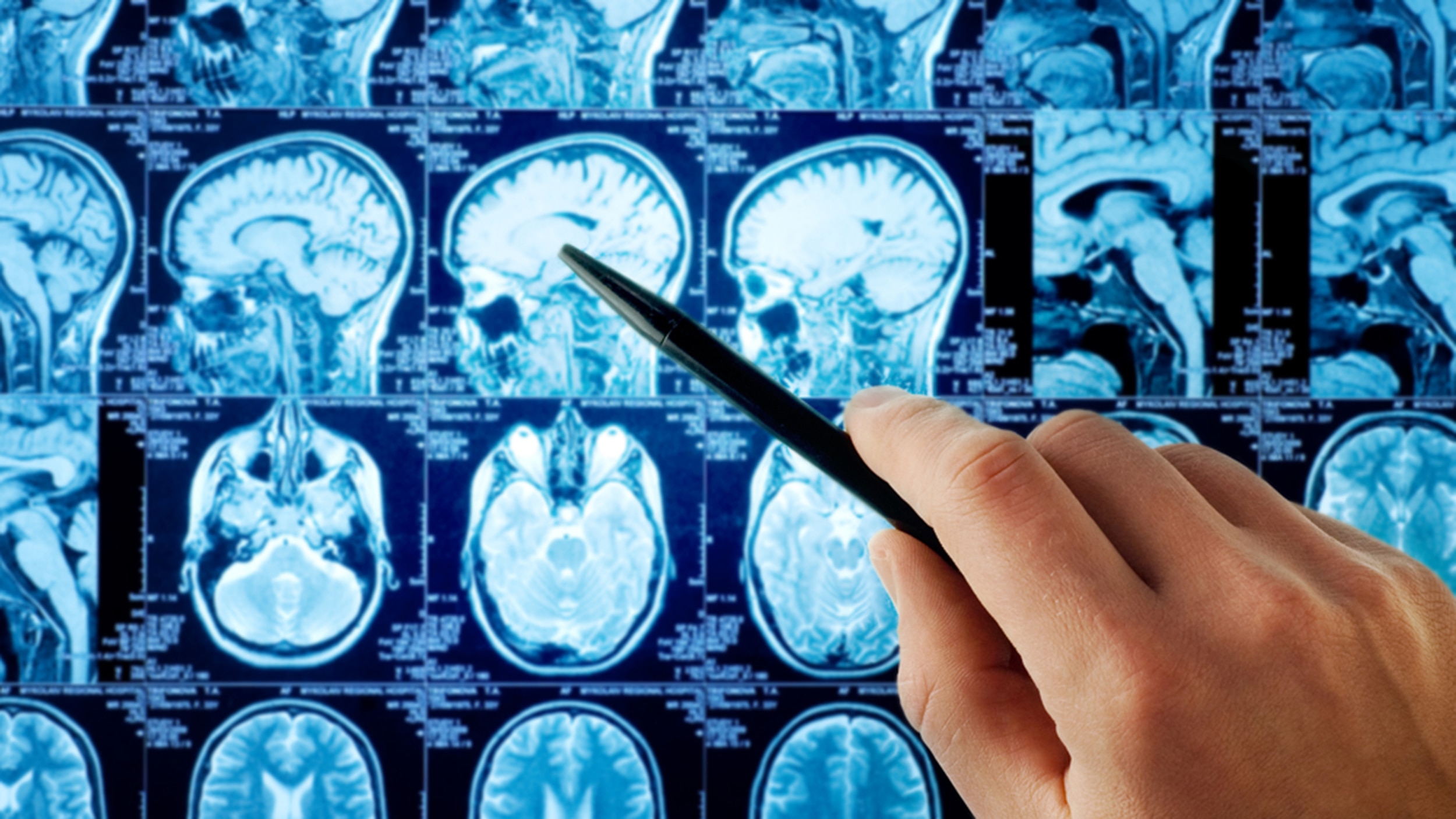 | Brain Surgery ₹1200 Brain tumours are abnormal growths of cells within the brain or the surrounding structures. View service → |
Brain Surgeon Here are numerous types of brain surgery. The type used is based on the area of the brain and the condition being treated. View product → |
What should happen after surgery, and what does pathology change?
After the operation, pathology examines the tissue and, when indicated, molecular testing identifies the tumor’s exact type, grade, and relevant markers. These results can alter prognosis and determine whether you need radiotherapy, chemotherapy, targeted treatment, or observation.
A postoperative MRI checks for residual tumor and establishes a new baseline. Scheduled surveillance scans then monitor recurrence. An apparently complete resection may not cure an infiltrative tumor because microscopic cells can extend beyond the visible MRI abnormality.
Recovery may include wound care, rehabilitation for weakness, speech, vision, memory, or mobility problems, and seizure management with medication and safety advice. Contact the treating team urgently for a new seizure, worsening weakness or speech, severe headache with repeated vomiting, reduced consciousness, fever with wound problems, or sudden neurological deterioration.
Use this checklist when reviewing your brain tumor surgery explained plan:
- Confirm the final pathology and molecular report.
- Ask when the postoperative MRI and next surveillance scans are due.
- Clarify rehabilitation goals, seizure treatment, and recurrence monitoring.
- Ensure the brain tumor consultation records the diagnosis, surgical objective, alternatives, patient-specific risks, recovery expectations, follow-up schedule, and triggers for urgent contact.
Frequently asked questions
When is brain tumor surgery needed?
Surgery is considered when a tumor causes symptoms, seizures, pressure, swelling, or neurological problems, or when tissue is needed for a diagnosis. Observation or non-surgical treatment may be safer for selected tumors.
What happens during a brain tumor consultation?
The surgeon reviews when symptoms began, how they changed, and whether seizures occurred. They perform a neurological examination and assess MRI scans, often with and without contrast, before discussing biopsy, removal, and treatment goals.
How do surgeons choose between biopsy and tumor removal?
The choice depends on the tumor’s location, size, appearance on MRI, symptoms, surgical risk, and whether a tissue diagnosis will change treatment. Removal is considered when safe, while biopsy is used when sampling provides information with less risk.
How should you prepare for a brain tumor consultation?
Bring MRI images and reports, previous medical records, a list of medicines and allergies, seizure or symptom details, and questions about diagnosis, surgical goals, risks, recovery, and alternatives.
What does pathology change after brain tumor surgery?
Pathology identifies the tumor type and grade and can guide additional testing, radiation, chemotherapy, targeted treatment, follow-up scans, and referrals to other specialists.
Related items
| TUMOR & CANCERS Brain Tumor & Cancer An Cerebral aneurysm or intracranial aneurysm is when the internal walls of the artery through which the blood flows in the brain becomes thin and... |
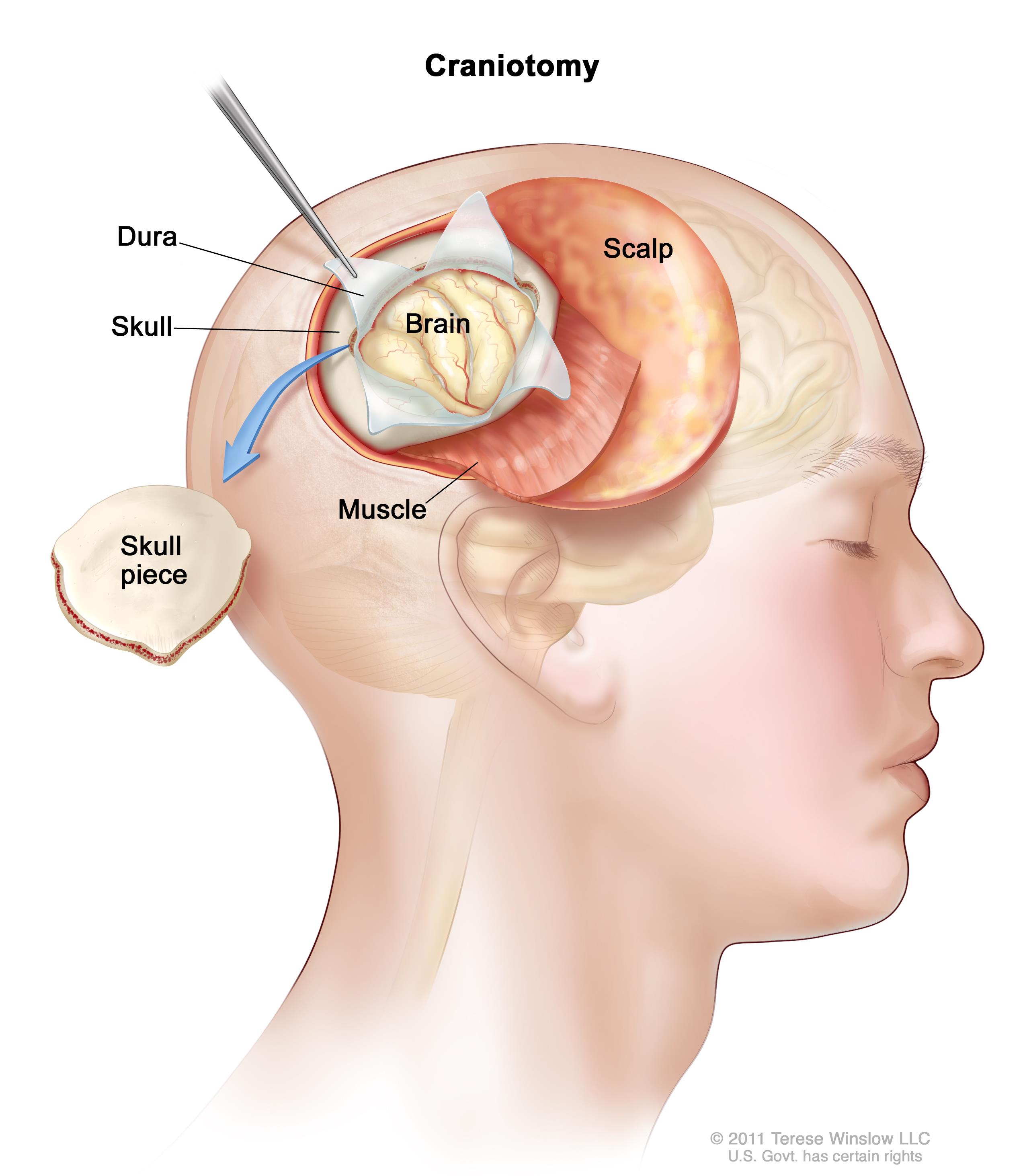 | Types of brain tumours- Primary brain tumors are tumors that originate in the brain itself, as opposed to secondary tumors that spread to the brain... View service → |